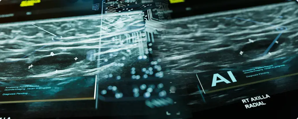
Can routine radiology workflows be faster, more accurate, and less burdensome? With the rise of artificial intelligence (AI), the answer is yes. As imaging volumes rise and staffing constraints tighten, AI is becoming a key tool in helping radiology teams manage and automate repetitive tasks with speed and consistency.
AI radiological applications now assist with case triage, image analysis, report generation, and other routine processes that often consume valuable time. By automating these tasks, radiologists can focus on high-complexity cases while maintaining quality and turnaround expectations. AI doesn’t replace clinical judgment. Instead, it supports it with reliable input that improves efficiency and diagnostic confidence.
This article explores how AI is streamlining routine radiology workflows. We’ll cover the most common AI-powered tasks, key benefits like faster reporting and lower error rates, and the limitations practices should be aware of. Along the way, we’ll reference real-world statistics and insights to offer a complete picture of AI’s growing role in radiology.
- AI-driven automation offloads repetitive tasks such as triage, measurements, segmentation, and initial reporting.
- Automated workflows improve radiology efficiency, turnaround times, and diagnostic consistency across high-volume settings.
- Integrated AI reduces burnout by minimizing manual work and allowing radiologists to focus on complex interpretation and patient care.
- Successful automation depends on high-quality data, seamless RIS/PACS integration, and proactive governance to address bias and adoption challenges.
The Need for Automation in Radiology
As imaging volumes continue to rise and radiology teams face growing pressure to deliver results faster, automation has become both a clinical and operational imperative. Radiologists are tasked with handling more demanding workflows amid staffing constraints and escalating burnout. By automating repetitive processes, AI helps sustain consistency and quality, even in high-pressure environments.
Rising Demand and Radiologist Shortage
Globally, radiologists are in short supply. The volume of imaging studies increases by up to 5 percent annually, while the number of trained radiologists is not keeping pace. In the United States alone, a shortage of up to 42,000 radiologists is projected by 2033. Across Europe, the UK’s NHS reports a 30 percent workforce shortfall, and some lower-income countries have fewer than two radiologists per million people. As the gap widens, radiology departments are under pressure to do more with fewer resources.
Time-Consuming Routine Tasks
Tasks like measurements, comparisons, image labeling, and report drafting demand time but do not always require high-level interpretation. Automating these repetitive steps helps radiologists prioritize complex cases and dedicate more attention to clinical decision-making. Solutions like OmegaAI® and PowerServer™ enable intelligent triage, automatic segmentation, and smart worklist management that route studies based on urgency, all of which contribute to faster and more efficient care delivery.
Burnout and Human Error
Burnout continues to affect over 45 percent of radiologists, driven by long hours and administrative burdens. Fatigue elevates the risk of human error, particularly during high-volume or overnight shifts. AI-powered image analysis assists radiologists by detecting critical findings, enabling prioritization, and supporting second-read workflows. When integrated thoughtfully, AI enhances diagnostic confidence and contributes to safer, more efficient care.
These advantages reflect the broader benefits of artificial intelligence in radiology, where image analysis and automation helps reduce diagnostic variability, accelerate workflows, and support high-quality, patient-centered care without compromising clinical oversight.
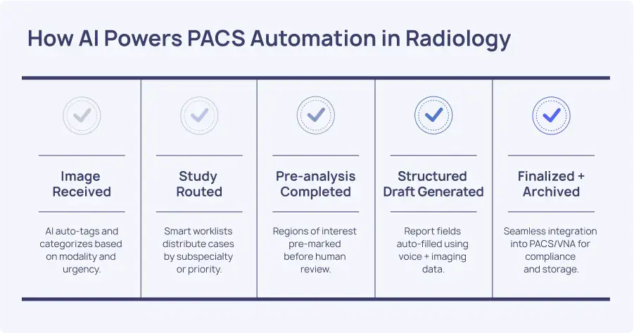
Automated Radiology Tasks Powered by AI
Artificial intelligence is transforming the daily workflow of radiology departments by automating routine, repetitive tasks that previously consumed valuable time and attention. AI enhances precision, accelerates case reviews, and facilitates radiology teams to dedicate more focus to high-complexity cases and clinical decision-making.
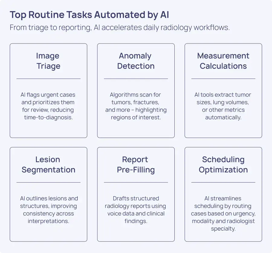
Image Analysis and Detection
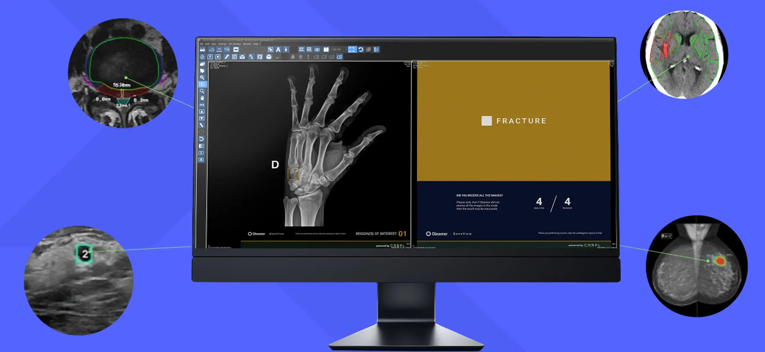
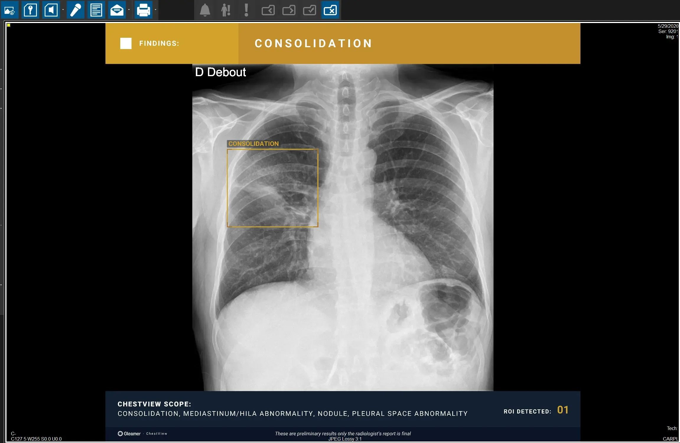
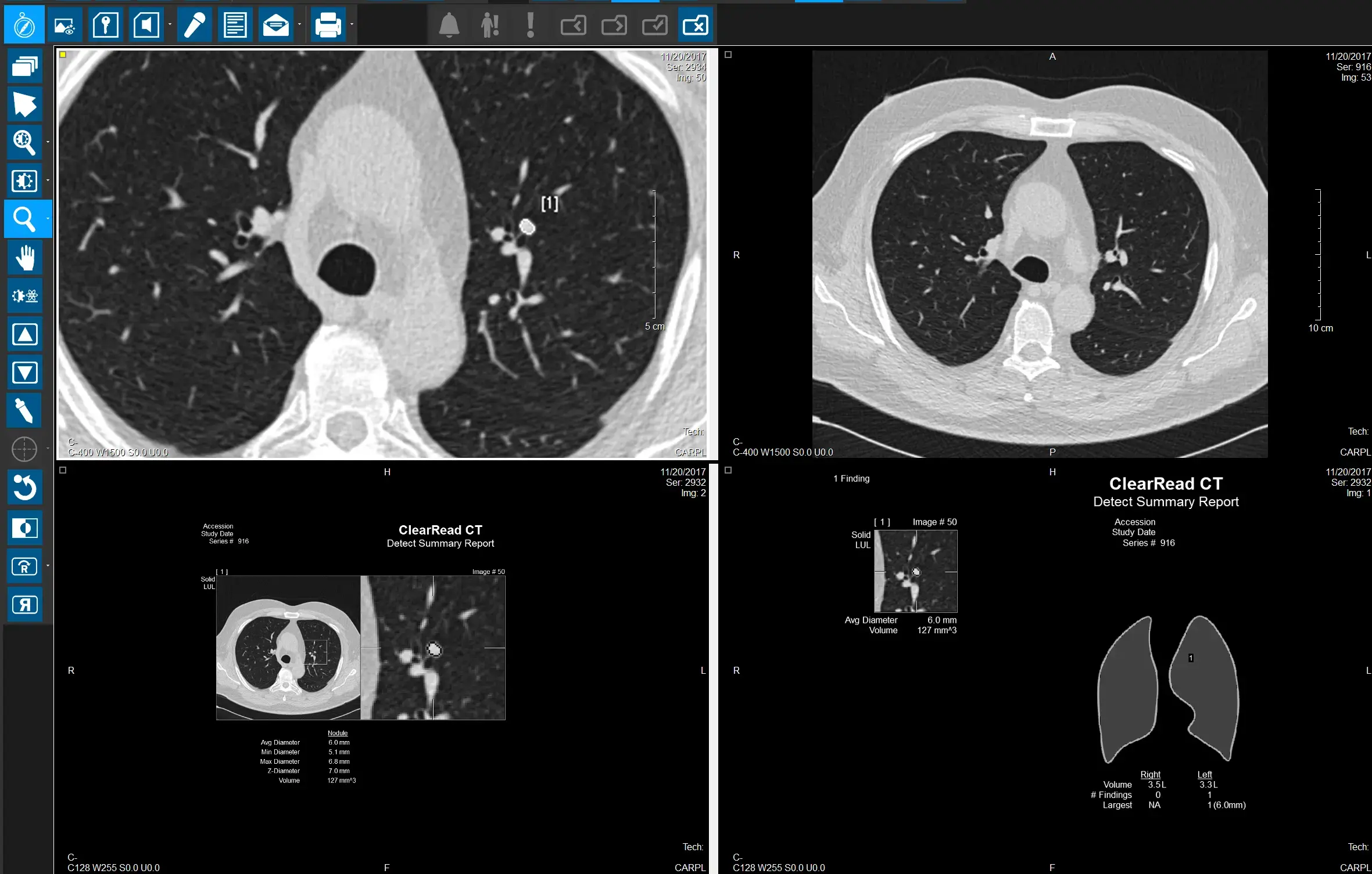
AI algorithms are trained on large volumes of annotated imaging data to identify patterns associated with abnormalities such as tumors, fractures, hemorrhages, and lung nodules. Using modalities like CT, MRI, and X-ray, these tools quickly analyze image sets and highlight regions of interest for further review.
By scanning thousands of studies with high accuracy, AI improves both speed and diagnostic consistency. Peer-reviewed studies report that YOLO-based models have achieved up to 98.7% accuracy in detecting lung nodules in CT scans[1]. This level of performance enables radiologists to focus more on advanced interpretation and differential diagnoses, contributing to stronger clinical outcomes and greater diagnostic confidence.
Workflow Triage and Case Prioritization
AI-enabled triage platforms analyze incoming imaging studies in real time, automatically classifying them based on urgency and clinical relevance. Critical findings such as suspected stroke, pneumothorax, or intracranial hemorrhage are prioritized and pushed to the top of radiologists’ worklists. Statuses can be customized by each facility to reflect local clinical protocols and priorities. These intelligent systems integrate directly into RIS, PACS, and enterprise imaging platforms to maintain seamless workflow continuity.
Such automation is particularly vital in emergency radiology and high-volume imaging centers, where delays in interpretation and interpretation and undertriage of high-risk conditions can delay urgent medical care. For example, AI-assisted chest X-ray interpretation has helped cut average turnaround times from 11.2 days to 2.7 days[2], illustrating the operational and clinical value of automated prioritization. This approach improves time-to-diagnosis, enhances radiologist productivity, and reduces length of stay for high-risk patients. AI in emergency medicine gives visibility to critical cases with worklist alerts of the most critical cases versus “first in, first read”.
Segmentation and Annotation
AI tools now handle tasks like lesion segmentation, automated anatomical labeling, and region-of-interest identification with impressive accuracy. These features reduce manual effort and improve the consistency of image annotations across radiologists and institutions.
Automated segmentation plays a critical role in surgical planning, radiation therapy targeting, and treatment response tracking. For instance, AI-enabled systems can calculate tumor volumes, mark anatomical landmarks, or track disease progression over time. This level of automation ensures reproducibility, supports precision medicine, and reduces variability in complex clinical workflows.
By standardizing annotations and minimizing manual input, AI also reduces inter-observer variability, a key factor in diagnostic agreement and longitudinal studies.
Report Generation and Documentation
AI-powered systems can extract structured data from imaging studies and generate draft reports for radiologists to review and finalize. Natural language processing (NLP) further enhances this process by converting medical findings into readable, concise summaries with less dictation.
NLP algorithms are particularly useful for transforming spoken or written notes into structured radiology reports. RamSoft’s OmegaAI® and PowerServer™ integrate voice recognition tools with structured reporting templates to auto-fill diagnostic impressions, measurements, and procedural details. AI processes also automate comparison and template selection, key findings dictation mapping, and automated billing code generation.
This reduces the cognitive load on radiologists and minimizes clerical errors.
Clinics adopting AI-assisted reporting tools have reported measurable gains in both speed and accuracy. Radiologists are able to finalize reports more efficiently, meet service-level agreements, and spend additional time communicating findings with referring physicians. In high-volume imaging environments, this level of automation significantly improves report turnaround and supports a more seamless continuum of care.
Benefits of AI for Efficiency in Radiology
Artificial intelligence improves more than just workflows. It helps radiology departments deliver faster, more consistent, and resource-conscious care. As patient volumes increase and teams are asked to do more with fewer resources, AI acts as a strategic tool that targets inefficiencies and enhances how imaging services are delivered. The following areas demonstrate how AI significantly contributes to operational efficiency in modern radiology settings.
Boosted Speed
AI improves scan efficiency by reducing the time between image acquisition and diagnosis, automating early steps in the interpretation process. Instead of reviewing every image manually, radiologists can rely on AI algorithms to pre-analyze studies, highlight key findings, and sort cases based on clinical urgency then route the study to the radiologists speciality. In busy hospital systems or teleradiology networks, this functionality is critical to maintaining service-level expectations.
AI also helps eliminate backlog by handling routine cases more quickly, improving overall patient throughput. Faster review times lead to shorter reporting windows and quicker treatment decisions, which are especially important in emergency and acute care environments.
Diagnostic Accuracy and Consistency
AI platforms are trained on vast datasets and follow consistent interpretive criteria, which helps standardize diagnostic performance across radiologists and shifts. These systems are particularly effective in identifying subtle patterns that may be overlooked in time-constrained environments, such as microfractures, early-stage tumors, or slight ischemic changes.
As part of AI diagnostics, these tools serve as a second reader—boosting confidence and reducing errors of omission during image interpretation.AI assists in overcoming reading biases such as biases influenced by history, satisfaction of search, and distraction caused by pathology.
This level of consistency is especially valuable in quality assurance programs and long-term disease monitoring, where accurate comparisons over time are essential.
Reduced Administrative Workload
AI significantly reduces the time spent on non-clinical tasks such as documentation and image labeling. Radiologists often devote considerable hours to administrative work that does not require clinical interpretation. With AI tools powered by natural language processing and voice recognition, much of this documentation can be automated. The solutions help extract relevant data from EMR notes, history, labs, surgical histories then, auto-populate templates, and draft report summaries. This allows radiologists to spend more time reviewing complex cases and consulting with referring physicians, improving both productivity and job satisfaction.
Cost Savings and Resource Optimization
AI enables radiology departments to improve efficiency without necessarily expanding staffing or infrastructure. By reducing turnaround times and increasing report accuracy, AI helps minimize the need for repeat scans and unnecessary follow-ups. These improvements translate into better use of scanners, more predictable scheduling, and a higher volume of studies per day.
Institutions that implement AI tools are better positioned to meet rising demand without sacrificing quality. For a deeper look at how this works in practice, explore the key benefits of artificial intelligence (AI) for healthcare imaging.
Limitations and Risks of AI in Radiology
Artificial intelligence is becoming an essential part of modern radiology workflows, offering meaningful gains in speed, accuracy, and efficiency. However, its implementation introduces new complexities that demand strategic consideration and strong governance. From data quality and system integration to regulatory concerns and clinician acceptance, these factors must be addressed to ensure safe and effective implementation across imaging departments.
Data Quality and Algorithm Bias
The reliability of AI models depends heavily on the quality and diversity of the data they are trained on. High-quality, diverse datasets are essential to building algorithms that perform reliably across a wide range of clinical scenarios. When training data lacks representation, models may produce biased or inaccurate results. This can lead to misdiagnoses or unequal outcomes for underrepresented patient populations.
Additionally, errors in labeled datasets or inconsistencies in annotations can misguide machine learning processes, reducing model accuracy. Radiology teams and AI developers must invest in robust data governance practices, including validation across multiple sites and patient groups, to improve generalizability and minimize bias. Continuous model retraining with new and verified data is also necessary to maintain clinical relevance and safety over time.
Integration into Existing Systems
One of the most overlooked hurdles in AI adoption is the technical complexity of integrating new tools into established radiology workflows. Many departments rely on legacy PACS, RIS, and EHR systems that are not inherently designed to support AI applications. Without a well-planned integration strategy, introducing AI can create friction and disrupt operational continuity rather than streamline it.
Successful deployment requires interoperability with existing systems and minimal disruption to the clinical workflow. AI tools must seamlessly plug into the radiologist’s environment, eliminating the need to switch between worklists and platforms or manually re-enter data. This is where robust IT infrastructure and adaptable software architecture are critical.
RamSoft’s PowerServer™ (a cloud-based RIS/PACS suite) and OmegaAI® (a zero-footprint, cloud-native RIS/PACS/VNA platform) are designed for seamless integration with any radiology infrastructure. Both support industry standards like FHIR, HL7, and DICOM, and easily connect with third-party tools such as RADPAIR,Remedy Logic, iCAD, CARPL, Therapixel, and NewVue; making them highly adaptable to diverse clinical environments without disrupting existing workflows.
For healthcare networks operating across multiple locations or specialties, OmegaAI’s cloud-native architecture further simplifies centralized integration and scaling. With configurable workflows and broad compatibility, RamSoft’s solutions empower IT teams and clinical leaders to adopt AI with confidence without compromising on security, data flow, or productivity.
Regulatory and Ethical Concerns
Using AI in a medical setting raises important regulatory and ethical issues. Patient data must be handled with strict safeguards to ensure privacy and compliance with healthcare laws such as HIPAA and GDPR. AI systems must be transparent in how they process data and generate outputs to maintain trust among clinicians and patients alike.
There is also a need for ethical oversight. Overreliance on automated tools without proper human review can lead to unintended consequences. Regulators, hospital boards, and developers must collaborate to set clear boundaries for how and when AI is used. This includes defining accountability when errors occur and establishing standards for algorithm validation before clinical deployment. Ongoing post-market surveillance is equally important to track real-world performance and address emerging risks.
Resistance to Adoption
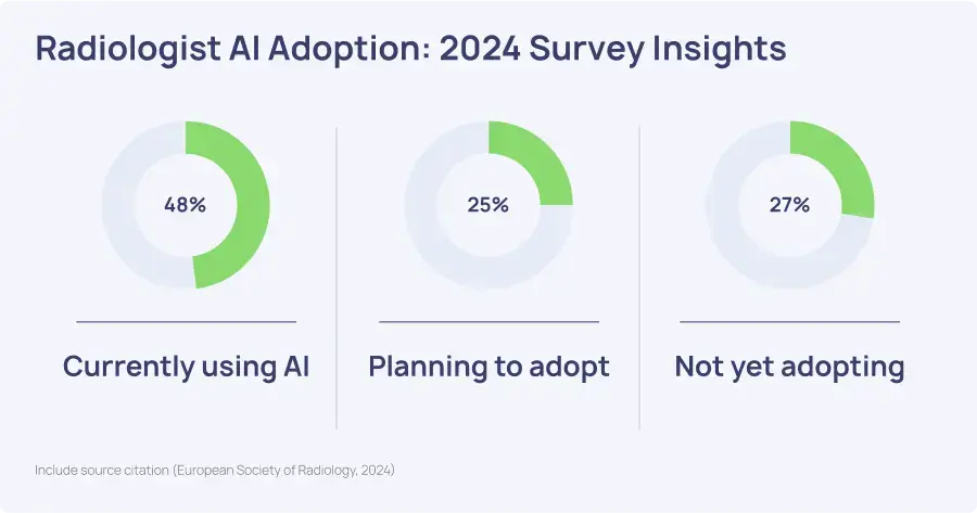
Despite promising advancements in diagnostic accuracy and workflow efficiency, resistance to AI remains strong among radiologists. In a 2024 survey, nearly half of respondents expressed concern that AI could eventually replace parts of their work. These fears are particularly prevalent among more experienced radiologists, who report lower readiness to adopt AI compared to their early-career counterparts.
Algorithm transparency and reliability are also persistent barriers. According to the same survey, 42.9 percent of radiologists questioned whether AI could be trusted in critical diagnostic situations.
The literature also emphasizes that lack of awareness, limited hands-on exposure, and absence of structured implementation strategies contribute to ongoing hesitation. Radiologists who have only basic knowledge of AI are significantly more likely to fear its integration, while those with advanced familiarity report greater confidence. This indicates that targeted education and early engagement are key to improving adoption and trust.
A collaborative approach can mitigate these concerns. When radiologists are involved in the evaluation and deployment of AI tools, they are more likely to view them as clinical aids rather than replacements. Integrating AI into the medical curriculum and continuing education programs not only addresses technical gaps but also reframes AI as a partner in care—one that enhances, rather than undermines, the radiologist’s role.
The Future of Automation in Radiology
AI's Role in Radiology
Artificial intelligence is transforming how radiologists work by handling repetitive tasks such as image analysis, segmentation, and structured reporting. These capabilities allow radiologists to concentrate on interpretation, patient care, and clinical decision-making. AI serves as a trusted partner, helping to improve diagnostic consistency, reduce fatigue, and support timely, high-quality care.
By prioritizing urgent studies, extracting measurements instantly, and organizing data efficiently, AI enhances both speed and accuracy in the imaging workflow. This partnership leads to improved reporting quality and shorter turnaround times. As radiologists face increasing imaging volumes and complexity, AI provides the support needed to keep pace without compromising clinical standards.
Looking Ahead to AI Radiology
The future of AI in radiology points toward more intelligent, connected, and context-aware systems. As technology evolves, AI will increasingly combine medical imaging with other clinical data sources to offer a more complete diagnostic picture. This level of integration will help radiologists make even faster, more informed decisions in real time. AI benefits patients by shortening scan times, reducing radiation. In addition, AI is being developed to reduce or omit contrast media particularly in MRI and CT by generating synthetic post-contrast images through AI computational modeling.
Continued growth will rely on collaboration across healthcare, from radiologists and IT leaders to regulatory bodies and developers. Setting clear standards for safety, transparency, and clinical validation remains essential. In the broader future of AI in radiology, automation will not only boost efficiency but also strengthen patient care through better coordination, reduced delays, and more confident diagnoses.
Ready to See AI in Action?
Discover how RamSoft is helping radiology practices thrive in the future of AI in radiology. Our cloud-native RIS, PACS, and VNA solutions are designed to accelerate imaging workflows, improve diagnostic confidence, and integrate seamlessly with the tools you already use. With built-in AI functionalities and endless integration possibilities, you can streamline operations, enhance collaboration, and deliver exceptional care with confidence.
Request a demo today and explore what intelligent automation can do for your practice.
Frequently Asked Questions
Can radiology be automated?
Yes, many aspects of radiology can be automated using artificial intelligence. While fully replacing the radiologist is neither the intent nor the reality, automation supports key tasks such as image triage, anomaly detection, measurements, report generation, and follow-up recommendations. This enables radiologists to dedicate more attention to high-value tasks, improving both efficiency and diagnostic confidence.
What are the benefits of automating radiology tasks?
Automating radiology tasks offers clear operational and clinical benefits. It reduces turnaround times, minimizes human error, and increases reporting consistency. Radiologists spend less time on repetitive work like measurements and protocoling, and more time on complex diagnostic decisions. Automation also helps manage rising imaging volumes, reduces fatigue, and improves overall workflow efficiency. Ultimately, patients receive faster, more accurate results with fewer delays.
What can AI do in radiology?
AI imaging solutions can analyze medical images, detect abnormalities, segment structures, prioritize urgent cases, and assist with generating structured reports. It enhances diagnostic precision by uncovering subtle indicators that may not be easily noticeable during manual review. AI can also flag inconsistencies, streamline documentation, and reduce administrative overhead.
What types of radiological tasks can AI automate?
Radiology task automation covers image triage, anomaly detection, measurement calculations, report pre-filling, and follow-up recommendations. It also supports workflow automation by organizing study priorities, optimizing scheduling and front desk operations. These automated functions help radiology departments increase throughput, standardize outputs, and improve diagnostic consistency across high volumes of imaging data.
How many radiologists use AI?
AI adoption in radiology is accelerating. According to a 2024 survey by the European Society of Radiology, 48% of radiologists are already using AI tools in clinical practice, with another 25% planning to adopt them soon. Usage is especially high in large hospitals and teleradiology groups. As trust in AI grows, adoption is expected to expand across healthcare settings of all sizes, supporting the future of AI in radiology.
References
[1] Abdulqader, A. F., Abdulameer, S., Bishoyi, A. K., Yadav, A., Rekha, M. M., Kundlas, M., Kavitha, V., Aminov, Z., Abdulali, Z. S., Alwan, M., Jawad, M., Mushtaq, H., & Farhood, B. (2025). Multi-objective deep learning for lung cancer detection in CT images: Enhancements in tumor classification, localization, and diagnostic efficiency. Discover Oncology, 16, 529. https://doi.org/10.1007/s12672-025-02314-8
[2] Irmici, G., Cè, M., Caloro, E., Khenkina, N., Della Pepa, G., Ascenti, V., Martinenghi, C., Papa, S., Oliva, G., & Cellina, M. (2023). Chest X-ray in emergency radiology: What artificial intelligence applications are available? Diagnostics, 13(2), 216. https://doi.org/10.3390/diagnostics13020216